Preclinical Imaging
Overview
Our diverse team of experts have experience in instrument engineering, signal processing, and pharmaceutical R&D, collectively resulting in a long history designing and executing imaging and pathology studies. Our scientists have performed hundreds of studies across multiple therapeutic research areas, including oncology, neuroscience, inflammation, cardiovascular, metabolic, and musculoskeletal, and our experience runs the gamut of applications. We understand the physiological and biochemical mechanisms that affect imaging—targets, models, kinetics, and more. This vast hands-on experience allows us to understand drug behavior and translate knowledge to provide effective studies and useful data.
We define imaging as assays that enable visualization of subcellular distribution and processes all the way to translational and diagnostic clinical imaging biomarkers, including everything in between. With all imaging techniques, we use methods to ensure data is quantifiable and leverage our powerful analytics platform to ensure that results can be used to drive meaningful decisions.
Learn more – Radiopharmaceutical therapy
Infographic – Radiopharmaceutical Drug Development
Capabilities
Execution of these studies requires the skills of many scientific disciplines and the structure to enable the complex coordination of all facets of the experiment, including animal models, radiolabeled molecules, multiple timepoints, and multi-modal imaging. Invicro has built a research laboratory network with services that are necessary to support these imaging endpoints, and we continue to advance and add to these services to meet the needs of our sponsors. We often develop protocols for novel compounds, work through the radiochemistry needs together with the sponsor, and assist with animal model development as needed. We can perform studies on a range of different species with a large number of administration routes available. Species include rodent, dog, cat, pig, rabbit, sheep, and non-human primates as well as surgical and oncology models.
Key Supporting Capabilities |
Complete radiopharmaceutical and theranostics services |
Disease model development, including tumor inoculation |
Pharmacology, efficacy, and biomarker study execution |
Drug and imaging agent administration, including during scanning |
Blood collection for whole blood, plasma, or serum PK analysis |
Tissue collection and biomarker development |
Gamma and liquid scintillation counting (biodistribution studies) |
Focal beam radiation / Whole body irradiation |
Applications
Antibody-based Theranostics
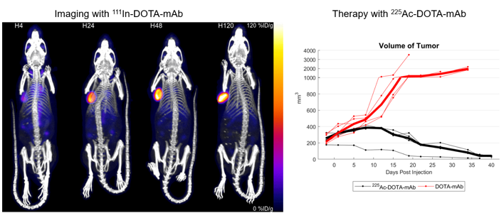
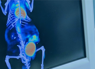
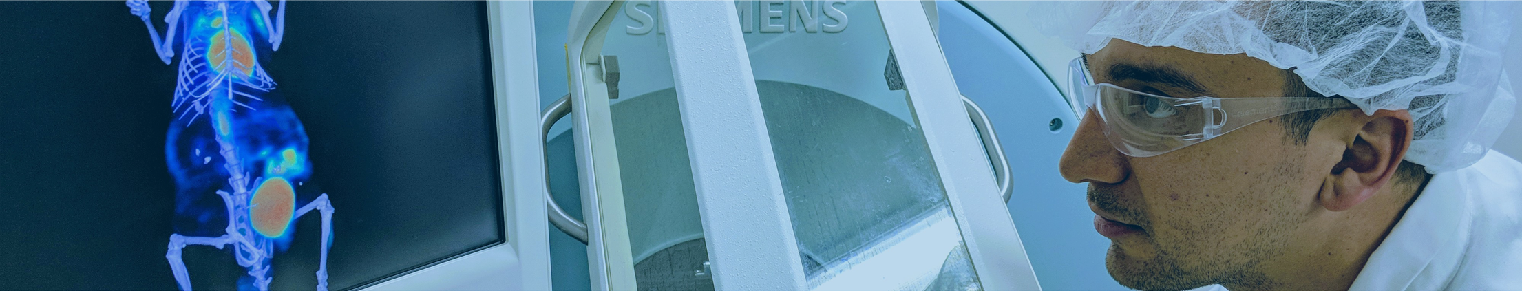
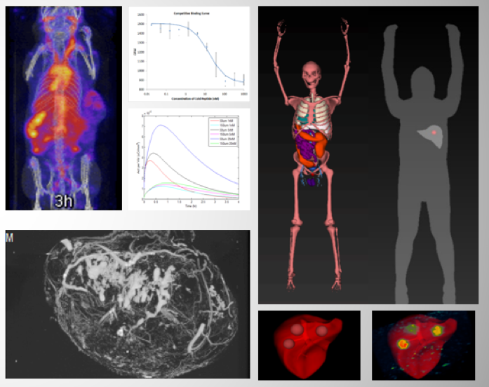
Invicro has extensive experience developing imaging solutions for antibody-based radiotherapies. In this example, imaging was performed using 111In while the antibody therapy utilized 225Ac. This imaging approach allows for the visualization of pharmacokinetic profiles of radionuclide therapies in vivo and confirms the recruitment of the targeted-therapy to the tumor.
Antibody-Drug Conjugate (ADC) Biodistribution
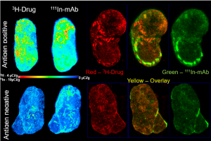
Invicro possess the expertise to image large molecule drugs, such as Antibody-drug Conjugates (ADCs). In this example, a dual-isotope quantitative 3D autoradiography method for simultaneous and quantitative assessment of both the antibody and drug conjugate distribution was used. This method allows for the tracking of the two major components of an ADC, the mAb and small molecule drug conjugate at the same time in three dimension over time.
Biodistribution of Radiolabeled Compounds
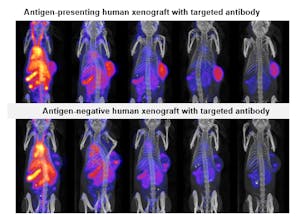
Invicro has run many biodistribution studies across a wide-range of therapeutic areas. In this study, targeted and control antibodies were radiolabeled for longitudinal SPECT/CT imaging to confirm tumor targeting, kinetics, and whole body biodistribution. To learn more, download the publication.
Candidate and Dose Selection Studies
Invicro can perform competitive tissue binding assays comparing labelled and non-radioactive drugs using a combination of in vitro assays and high-resolution ex vivo imaging techniques to confirm radioligand binding and inform dose selection for subsequent studies.
Determining Target Expression
Target densities (maximum binding signal, Bmax) and regional distributions may not be known. In order to determine these parameters, Invicro can utilize multiple approaches to measure target expression in ethically sourced animal and/or human tissue including:
- in vitro autoradiography
- Homogenate binding assays
- Bioactivity assays
- Internalization Assays
- in vivo Imaging
- FACS
- IHC
- Western Blotting
Drug Efficacy and Pharmacology

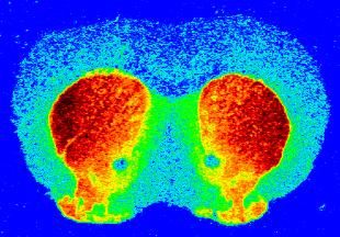
Invicro is a leading provider of optical imaging services to determine the efficacy of therapeutic candidates. In this example, efficacy of anti-tumor therapies targeting deep tissue tumor models (orthotopic) was determined non-invasively using Bioluminescence. This imaging approach allowed for efficacy to be determined with tissue relevant context. Invicro has experience working with a wide-range of tumor models including:
- Hematological
- Solid Tumors
- Metastatic
Dynamic MR Contrast Enhancement (DCE)
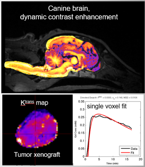 Dynamic contrast enhanced (DCE) T1-weighted MRI provides functional characterization of tumor perfusion and permeability, which may be heterogeneous. We have developed analysis tools for semi-automated estimation of parametric maps or ROI analysis.
High-Throughput Compound Screening
Invicro has extensive experience running high-throughput biodistribution studies in preclinical animal models. This service allows for the following experimental parameters to be determined:
- Whole body biodistribution
- Kinetics
- Off-target accumulation
Labeling of Drugs and Biomarkers
Invicro has extensive experience labeling different classes of molecules for in vivo and ex vivo imaging applications. From novel radioligand labeling using a risked-managed approach to meet specific customer needs, to GMP-based labeling for clinical imaging, the Invicro Chemistry team has the expertise required to label imaging agents using long and short-lived half life radionuclides for SPECT and PET along with fluorescence for optical imaging applications. We have direct experience with labeling the following types of molecules:
- Small Molecules
- Antibodies, Antibody Fragments
- Cells (T-cells)
- Viruses
- Liposomes
- Exosomes
- Nucleic Acids (ASO, RNA, DNA)
- Novel ligands
Metabolite Analysis by MRS
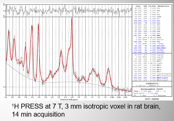 Invicro provides single voxel, in vivo 1H MRS. This technique can be used for longitudinal monitoring and therapy response of various metabolites and processes in parallel, for example, neurochemicals (Glu/Gln and GABA) and energy metabolism (PCr, lactate, and ATP). This method is also translatable to clinic. Invicro has various pulse sequences available (PRESS, semiLASER, ISIS, and STEAM) in addition to an automated LC Model (LCModel and Provencher) analysis workflow. Other applications are tissue pH (intra- and extracellular by P-31), quantification, distribution, and metabolism of exogenous F-19 labelled compounds as well as high-resolution ex vivo MR spectroscopy of tissue extracts.
Monitoring Gene Expression

Invicro’s cryo-fluorescence tomography (CFT) is a 3D, high resolution imaging modality that can track the biodistribution of various test articles, including gene and cellular therapies delivered using viruses and lipid nanoparticles. Registration to white light images and fly through movie generation provides accurate localization of signal. Visualization of anatomical details with high soft tissue contrast using in vivo MRI can evaluate morphological changes in brain, tumors, and more and detect small lesions (e.g., in the lung). The spatial resolution can be tailored as needed, as in the example below, for details such as optical nerves. Invicro can employ all standard MR contrasts and has access to various RF coils for whole body or body parts, with gating for improved spatial resolution and customizable analysis. CT and ultrasound are also available for structural imaging.
Modeling to Inform Clinical Protocol Design
 Combine preclinical discovery work with distributed tumor model simulations to directly guide clinical imaging protocols to support the estimation of tumor properties in human subjects and guide the design of first-in-human oncology clinical imaging protocols with novel biomarkers.
Pharmacological MR Imaging (phMRI)
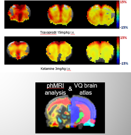 Quantifying changes in BOLD. This allows in vivo, longitudinal assessment of non-labelled compounds. Various mathematical modelling and simulation methods are available. Brain atlases are available for various species, including rodents and non-human primates.
Pharmacokinetic Modeling
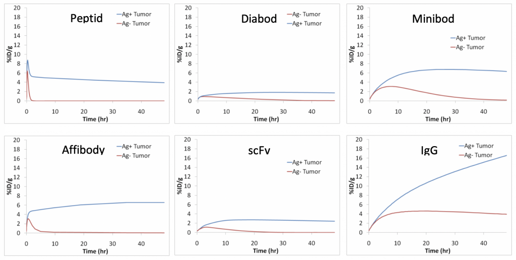 Invicro uses pharmacokinetic modeling to support study design and interpretation of experimental data. Invicro often provides PK modeling support in an iterative fashion to continually learn and modify the model and/or model parameters to build confidence in simulations to support experimental study design and clinical translation. The figure uses PK modeling to demonstrate the impact of molecular weight on tumor accumulation while keeping all other model parameters constant.
Radiopharmaceutical Drug Development
Bringing new therapeutic radiopharmaceuticals to the market is a complex and challenging process. When filing an Investigational New Drug (IND) or Clinical Trial Application (CTA) prior to the initiation of human trials, it is crucial to have strong non-clinical data to support a new candidate. Invicro provides complete preclinical support of your project, including:
- Study design and consultation
- Radiolabeling/conjugation to support alpha, beta, and gamma emitting isotopes
- in vitro/ex vivo cell and tissue-based assays
- Primary pharmacology studies to demonstrate MOA
- in vivo Imaging model development to support theranostic imaging
- Advanced image analytics
- Dosimetry
- IND support
- Development of chemistry, manufacturing and controls package for imaging agents
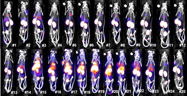
Screening Gene Delivery in vivo with Bioluminescence Imaging
Bioluminescence imaging (BLI) using luciferase reporters has revolutionized discovery research and is now a mainstay for in vivo testing in oncology (e.g., oncolytic virus and immune cell tracking) and for evaluating gene therapies delivered using lipid nanoparticles, viruses, and siRNA. In this example, different mRNA gene products were evaluated with or without supplemental test agents to determine effectiveness in promoting expression.
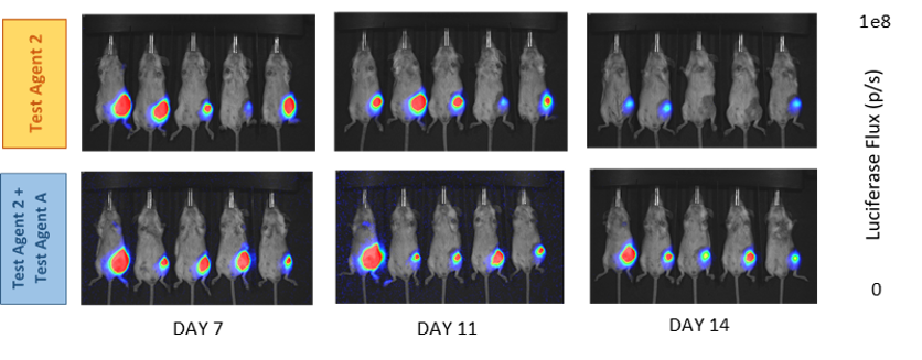
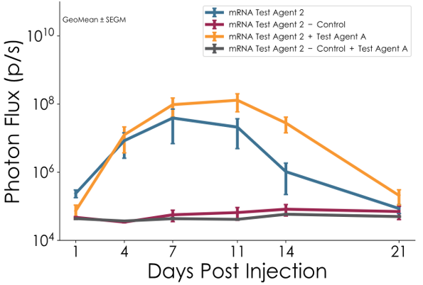
Gene delivery agent screening study: in vivo (italics in vivo) mRNA gene delivery time course for multiple test agents (route: IM) quantified by bioluminescence imaging. The graph shows hindlimb photon flux (p/s) vs. time, with the data plotted as geometric mean +/- standard error in geometric mean. Example images highlight a combination treatment benefit vs. Single agent for test Test Agent 2.
Structural/Anatomical Imaging
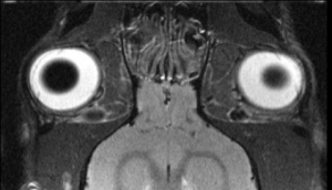 Visualization of anatomical details with high soft tissue contrast using in vivo MRI can evaluate morphological changes in brain, tumors, and more and detect small lesions (e.g., in the lung). The spatial resolution can be tailored as needed, as in the example below, for details such as optical nerves. Invicro can employ all standard MR contrasts and has access to various RF coils for whole body or body parts, with gating for improved spatial resolution and customizable analysis. CT and ultrasound are also available for structural imaging.
Tissue Microstructure
Diffusion-based MRI can be used to localize brain damage and monitor disease progression monitoring. Modelling for quantitative parametric maps is available.
Vascular Imaging (In Vivo and Ex Vivo with AltaBlu)
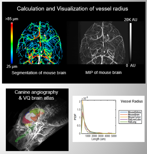 AltaBlu Prime is a proprietary radiopaque vascular casting agent used to image vasculature using microCT and 3D vasculature in whole animal and organ level angiogenesis in tumors and implants. Obtain segmentation, calculation, and visualization of vessel and radius and density. View a deeper branch-by-branch analysis of branch radius, length, vessel branching fraction, and tortuosity with characterization of vessel properties
Modalities
Invicro is imaging modality agnostic and can apply techniques across therapeutic areas and species. Studies may be single or multi-modal and may combine multiple isotopes or contrast mechanisms. When designing any study, we combine imaging theory, published methods, and the technical and professional expertise of our staff to ensure the appropriate technique is applied to best answer the biological question.
in vivo |
Tissue/Cell-Based |
PET |
Quantitative Whole-Body Autoradiography |
SPECT |
Microautoradiography |
MRI/MRS |
Optical 2D and 3D cryotomography |
CT |
Cryofluorescence Tomography (CFT) |
Bioluminescence |
Routine and Specialized Histology |
Fluorescence |
Immunohistochemistry (IHC) |
Ultrasound |
Blood and urine analysis |
Optoacoustic Imaging |
Flow Cytometry (FACS) |
Radioligand binding assays (cells and tissues) |
Therapeutic Areas
We specialize in the application of imaging in drug research and have experience working in multiple therapeutic areas. Our mission is to be a scientific partner to our sponsoring companies who benefit from our services for the rapid collection of data and advancement of their programs.

Drug Phases
Invicro’s discovery research team can provide guidance on all stages of the early drug development process from the choice of labeling technique, most appropriate imaging modality, choice of animal model, finalized study design, and preferred analysis endpoints to answer biological questions and produce meaningful results. Invicro has multiple sites globally with preclinical imaging facilities in Boston, Michigan, San Diego, New Haven, and London, UK and the site of choice will be dependent on sponsor needs.